| |
Anatomy images and medical illustrations
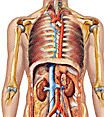
Medical illustration is the practice of depicting the human body and its structures to support teaching, study and clinical communication. Because the body's interior cannot easily be observed directly, accurate images are central to learning anatomy. They range from carefully drawn diagrams that label organs and tissues to detailed paintings that show the layered relationships between bones, muscles, nerves and blood vessels. A good illustration simplifies and highlights, drawing attention to the features a learner needs to grasp.
Anatomical images cover the whole body and its systems. Some depict the skeletal framework and the muscles, including the facial muscles and the nerves that control them; others show the heart and circulation, the brain and nervous system, or the organs of digestion and respiration such as the larynx. Regional illustrations focus on a particular area — the skull, neck, shoulder, hand and wrist, or the pelvis — while specialised images cover topics such as the reproductive organs, pregnancy and embryonic development, and the structure of sense organs like the eye and ear.
Modern study combines traditional illustration with images produced by medical technology. Photographs, microscope views of tissues and cells, and scans from techniques such as X-ray, CT and MRI reveal the real appearance of internal structures, complementing the clarity of a drawn diagram. Together these visual resources make anatomy easier to understand than text alone, helping students connect named structures with their actual shape, position and relationships within the body.
Frequently asked questions
- Why are illustrations important for learning anatomy?
- The body's interior cannot easily be seen directly, so clear drawings and diagrams help learners connect named structures with their shape, position and relationships.
- What do anatomical images usually show?
- They depict the body's systems and regions, from the skeleton and muscles to organs, sense organs and developmental stages such as the embryo.
- How do medical scans differ from drawn illustrations?
- Scans such as X-ray, CT and MRI capture the real internal appearance of a particular body, while drawings simplify and highlight features to aid understanding.
|
|
Human
anatomy images  related subject: Medical images
 |
| 3D
anatomy for medical students complex 3-D anatomical relationships |
|
Anatomical drawings blood vessels and lymph nodes, brain and spinal cord, digestive system,
epithelium: surfaces of the body, head and neck, heart-lung connections,
muscle-bone connections, urinary system |
| Anatomy and medical illustrations
atlas of the body, human atlas, anatomy, body, torso, brain, circulatory system,
circulation, digestive system, Endocrine System, ear, endocrine system, female
reproduction, reproductive, organs, pregnancy, hand, carpal tunnel, respiratory
system, respiration, muscles, nerves, nervous system, skeleton, skull, bones,
skin, urinary system |
|
Anatomy and medical illustrations
Body/Torso, Brain, Ear, Endocrine System, Eye, Female Reproductive System, Foot,
Hand, heart and Circulatory System, Lungs and Respiratory System, Lymphatic
System, Male Reproductive System, Muscles, Nervous System, Anatomy
Illustrations, Skeleton, Skin, Stomach and Digestive System, Teeth, Urinary
System, Educypedia |
|
Anatomy images 4 corps humain, en Français |
| Animating the
visible human data set quicktime movies |
|
Anterior and Posterior Views of Skull,
Lateral View of Skull, Sagittal Section Through Skull, Muscular Anatomy of Head
and Neck, Cerebral Hemispheres, Vertebral and Intervertebral Disc Anatomy,
Nervous System of Thorax and Upper Limb (Anterior View), Musculas Skeletal
Anatomy of Wrist and Hand (Palmar View), Viscera of Thorax (Anterior View),
Anatomy of the Heart (Anterior View), Abdominal Viscera (Anterior View),
Abdominal Viscera (Posterior View), Male Urogenital System (Midsagittal View),
Female Urogenital System (Midsagittal View), Nervous System of Pelvis and Lower
Limb (Anterior View), Muscular and Skeletal Anatomy of Ankle and Foot (Anterior
View), Skeletal Anatomy (Anterior View), Skeletal Anatomy (Posterior View),
Muscular System (Anterior View), Muscular System (Posterior View), Spinal and
Cranial Nerves, Arterial System (Anterior View), Venous System (Anterior View),
Lymphatic System (Anterior View), Respiratory System (Anterior View), Digestive
System (Anterior View), Urinary System (Anterior View) |
| Biotechnology
images from gene to function, biological engineering, cell processes,
building blocks, genetics, body atlas, chromosomes and cell division, viruses |
|
Color images of histological sections |
| Digital
anatomist interactive atlases movies |
|
Graphics Gallery is a series of labeled diagrams with explanations
representing the important processes of biotechnology. Each diagram is followed
by a summary of information, providing a context for the process illustrated |
| Guided tour visible human
18000 digitized sections of the body |
|
Human Biology |
|
Printable blank outline pictures of the human anatomy eye, ear, skin,
digestive system, respiratory system, circulatory system |
| Scanning
electron micrograph images of bacteria, viruses, insects and human cells |
| Visible
human cross sections |
|
5 senses pdf file |
|
Blood
circulation |
| Brain atlas java
based, structure and function, cerebrovascular disease, neoplastic disease (brain tumor),
degenerative Disease, inflammatory or infectious disease |
| Brain
connection graphics, images, audio, video and other material |
|
Cardiac muscle images |
| Cardiothoracic
imaging comprehensive overview of cardiothoracic imaging and major diseases, including over 700 diagnostic images, 100 original medical illustrations, and many animations and audiovisuals. Designed primarily for second-year medical students at Yale, the site may also be useful to science teachers, pre-med undergraduates, and anyone interested in the details of heart and lung imaging |
| Cells |
|
Cell: the virtual cell an
interactive, animated exploration of the cell |
|
Cytogenetics image gallery cytogenetics is the study of chromosomes and
chromosome abnormalities. The purpose of this Cytogenetics Gallery is to give
you the opportunity to see what chromosomes look like under the microscope and
how abnormalities of chromosomes are identified |
|
DNA from the beginning
animation, image gallery, video, problems, biographies, genes |
|
Ear |
|
Ear |
|
ECG image
index |
|
EKG library
an electrocardiogram (ECG / EKG) is an electrical recording of the heart and is
used in the investigation of heart disease. This library is a collection of
realistic looking recordings which will help improve your ECG skills |
|
Embryo: the human embryo
a three dimensional image reference of the human embryo based on magnetic
resonance imaging |
| Embryo images online |
|
Embryo images online |
|
Embryo: visible embryo
the spiral represents the 23 stages occurring in the first trimester of pregnancy and every two weeks of the second and third trimesters.
Use the spiral to navigate through the 40 weeks of pregnancy |
| Eye
cornea, aqueous, iris, lens, vitreous, retinal vessels, retina, macula, choroid, sclera, optic
nerve |
|
Eye & ear images |
| Hematopathology
index |
|
Histology atlas
Histology atlas |
|
Histology atlas
Histology atlas |
|
Histology atlas
Histology atlas, Cytology, Epithelium, Connective Tissue, Cartilage, Bone and
Bone Formation, Blood, Hematopoiesis, Muscle, Nervous Tissue, Cardiovascular
System, Lymphatic and Immune System, Integument, Upper Gastrointestinal Tract,
Lower Gastrointestinal Tract, Liver, Gall Bladder, and Pancreas, Respiratory
System, Urinary System, Endocrine System, Male Reproductive System, Female
Reproductive System |
|
Histology images
Peripheral Blood Cells, Tissues, Layers, and Organs, Epithelial Tissue, Surface
Specializations, and Glands, Connective Tissue, Cartilage and Bone and Bone
Histogenesis 20, cells of bone, Bone Marrow and Hemopoiesis, Nervous Tissue and
Neuromuscular Junction, Muscle Tissue, Integument, Circulatory System, Lymphoid
Tissues and Organs, Urinary System, Digestive System: Oral Cavity and Teeth,
Alimentary Canal, Liver, Gall Bladder, and Pancreas, Endocrine System,
Respiratory System, Male Reproductive System, Female Reproductive System |
| Molecular visualization freeware
software for looking at macromolecular structure and its relation to
function, rotating a protein or DNA molecule shows its 3D structure, a
tip |
|
Musculoskeletal
atlas |
|
Musculoskeletal system |
| Organ
system pathology images a
tip |
|
Sensory Systems Human Sensory Systems |
|
Skeleton |
|
Skeleton: bone
box |
|
Skeleton: ehe eskeletons project view the
bones of a human, gorilla |
|
Skeletal system allows you to access real photographs of the human skeletal
system |
|
Skeletal system |
|
Skeleton:
xray images of the human skeleton
the living skeleton: a tour of human bones |
|
Urinary
system jpg file |
|
Urogenital system real photographs of both male & female cat urinary &
reproductive systems, also included is a model of the kidney and real
photographs of a dissected kidney. |
|
Visible Human Female |
|
Visible Human Male
this coronal image is a reconstruction of the transverse images that were
captured to make up the Visible Human Male Database, ... |
|
|
Home
|
Site Map
|
Email: support[at]karadimov.info
Last updated on:
2026-06-24
|
Copyright © 2011-2021 Educypedia.
https://educypedia.org
|
| |
|
|